42 chlamydomonas diagram with labels
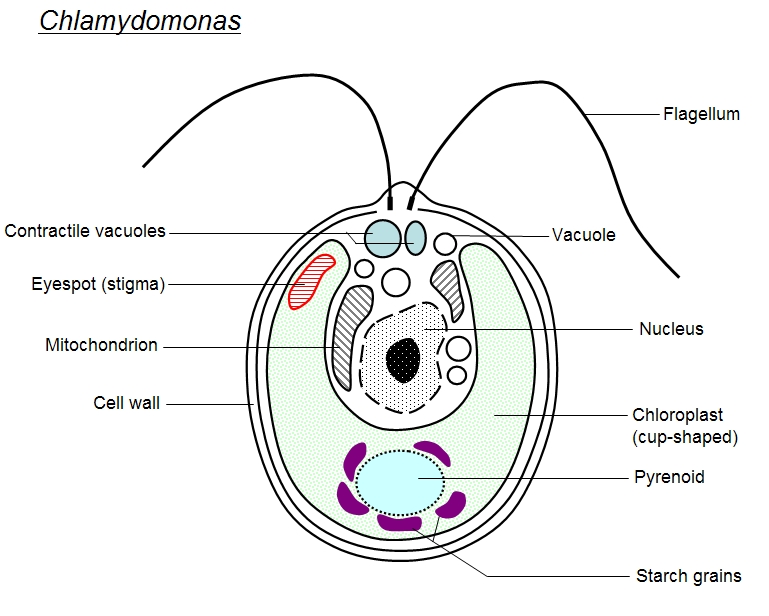
Labeled Diagram of Spirogyra - QS Study Labeled Diagram of Spirogyra Plant kingdom Spirogyra is a sophisticated, filamentous green alga, found in freshwater represented by about 300 species. It is also identified as pond silk, as its fiber burnishes like silk due to the occurrence of mucilage. The vegetative body structure of spirogyra A) External features Describe the structure of chlamydomonas with neat labelled diagram ... answeredOct 30, 2020by Naaji(56.8kpoints) selectedOct 30, 2020by Jaini Best answer 1. Chlamydomonas is a simple, unicellular, motile fresh water algae. They are oval, spherical or pyriform in shape. 2. The cell is surrounded by a thin and firm cell wall made of cellulose. 3. The cytoplasm is seen in between the cell membrane and the chloroplast. 4.
Eye Diagram With Labels and detailed description - BYJUS Well-Labelled Diagram of Eye The anterior chamber of the eye is the space between the cornea and the iris and is filled with a lubricating fluid, aqueous humour. The vascular layer of the eye, known as the choroid contains the connective tissue. The iris and the choroid are connected by the ciliary body.
Chlamydomonas diagram with labels
Chlamydomonas as a Model Organism - Rice University Chlamydomonas as a Model Organism. Chlamydomonas, a genus of unicellular photosynthetic flagellates, is an important model for studies of such fundamental processes as photosynthesis, motility, responses to stimuli such as light, and cell-cell recognition.C. reinhardi, the most commonly studied species of Chlamydomonas, has a relatively simple genome, which has been sequenced. Chlamydomonas Photos and Premium High Res Pictures - Getty Images Diagram of Chlamydomonas angulosa, Flagellated Protozoan. Drawing. Chlamydomonas, illustration. snow alga or snow algae -chlamydomonas nivalis-, kaunergrat range at the back, seeles see lake, kaunertal valley, tyrol, austria - chlamydomonas stock pictures, royalty-free photos & images. Draw a neat labelled diagram. Chlamydomonas - Biology Draw a neat labelled diagram. Chlamydomonas . Maharashtra State Board HSC Science (General) 11th. Textbook Solutions 8018. Important Solutions 19. Question Bank Solutions 5546. Concept Notes & Videos 439. Syllabus. Advertisement Remove all ads. Draw a neat labelled diagram. ...
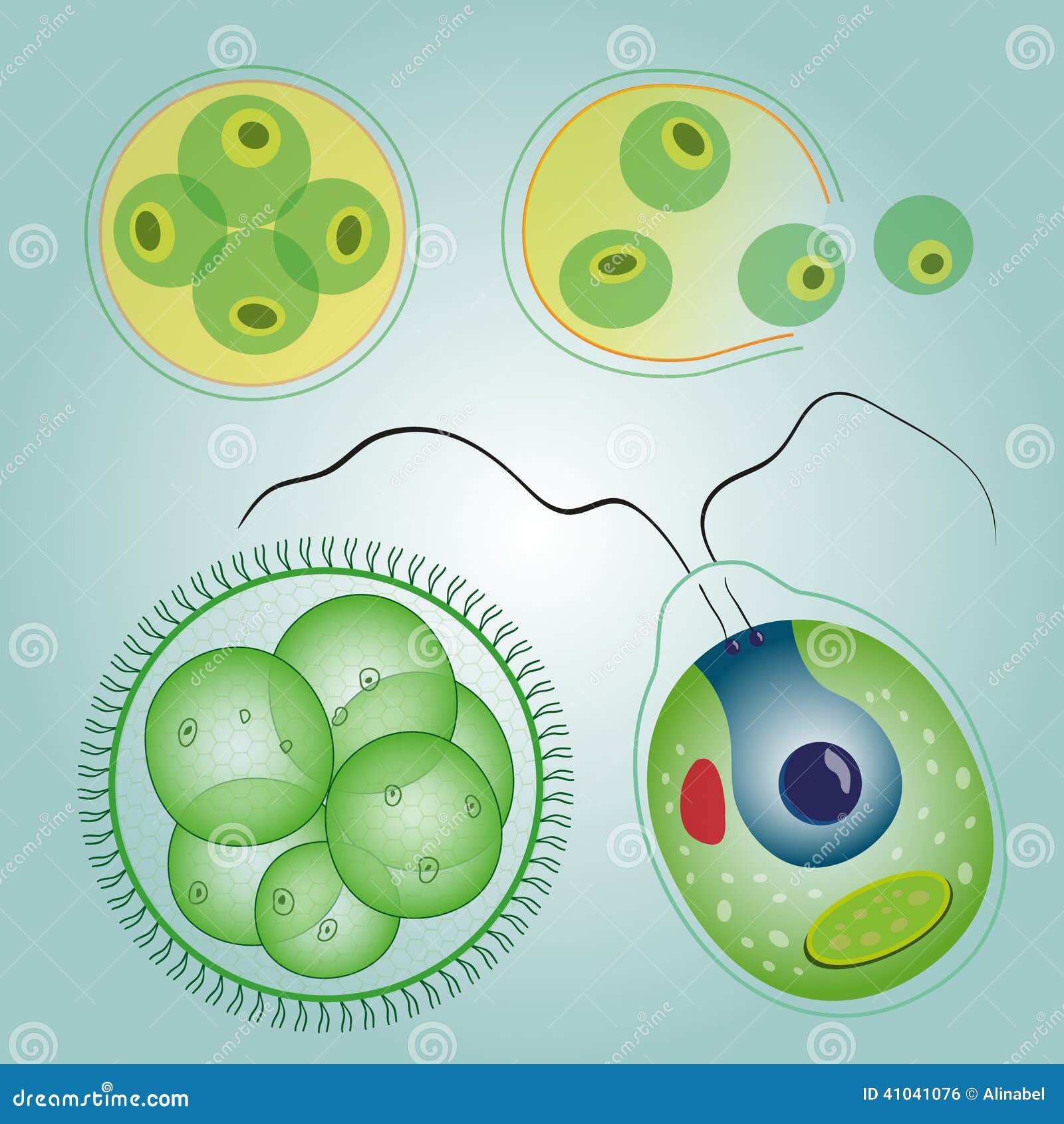
Chlamydomonas diagram with labels. Structure and Diagram of Volvox and Their Functions - NotesHippo Cell structure of volvox colony are Chlamydomonas type. Every cell has its own mucilage sheath (Fig. 1B). The mucilage envelope of colony appears angular due to compression between cells. The cells are connected to each other through cytoplasmic strands. In some species of Volvox the cytoplasmic connections or strands are not present. Chlamydomonas - Meaning, Structure, Life Cycle, Function and FAQs - VEDANTU Chlamydomonas is a model organism for molecular biology research, especially for the studies of flagellar motility, chloroplast dynamics, biogenesis, and genetics. Chlamydomonas have ion channels (channelrhodopsins) that are directly activated by light, which is one of its many distinguishing characteristics. Chlamydomonas | Cell wall, Single-celled organisms, Algae - Pinterest To sort things into groups or categories is a uniquely human characteristic. This hub looks at the defining features of the five kingdoms of life: Prokaryotae, Protoctista, Fungi, Animalia and Plantae. Structure of Chlamydomonas (With Diagram) | Genetics - Biology Discussion In this article we will discuss about the structure of chlamydomonas (explained with labelled diagram). The unicellular green alga Chlamydomonas is haploid with a single nucleus, a chloroplast and several mitochondria (Fig. 9.3). It can reproduce asexually as well as sexually by fusion of gametes of opposite mating types (mt + and mt - ).
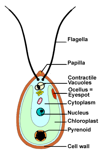
Spirogyra Labelled Diagram Draw a neat diagram of Spirogyra and label the following parts: i. Outermost layer of the cell. ii. Organelle that performs the function of. Each cell of Spirogyra filament is cylindrical and consists of 2 parts: cell wall and protoplast. The cell wall surrounds the protoplast, is protective and consists of. Clear Labeled Diagram Of Volvox - A Rubisco Binding Protein Is Required ... 12.10.2021 · labeled in the chlamydomonas diagram. Mean that a chlamydomonas is primitive itself. Volvox, chlamydomonas, and the evolution of multicellularity. The mucilage envelope of colony appears angular due to compression between cells. The cells are connected to each other through cytoplasmic strands. Animal Cells: Labelled Diagram, Definitions, and Structure - Research Tweet Only present in lower plant forms (e.g. chlamydomonas) Present in all animal cells: Chloroplast: Plant cells have chloroplasts to synthesize their own food. Absent: Plasma Membrane: Cell wall and a cell membrane: Only cell membrane: Flagella: Present in some cells (e.g. sperm of bryophytes and pteridophytes, cycads and Ginkgo) Chlamydomonas: Position, Occurrence and Structure (With Diagrams) Chlamydomonas is unicellular, motile green algae. The thallus is represented by a single cell. It is about 20 p,-30|i in length and 20 µ in diameter. The shape of thallus can be oval, spherical, oblong, ellipsoidal or pyriform. The pyriform or pear shaped thalli are common, they have narrow anterior end and a broad posterior end (Fig. 1).
Structure of Chlamydomonas (With Diagram) | Chlorophyta In this article we will discuss about the structure of chlamydomonas with the help of suitable diagrams. Chlamydomonas is unicellular, motile green algae. The thallus is represented by a single cell. It is about 20 p,-30|i in length and 20 µ in diameter. The shape of thallus can be oval, spherical, oblong, ellipsoidal or pyriform. HAP2 is essential for membrane merger in Chlamydomonas. (A) The plasma ... Download scientific diagram | HAP2 is essential for membrane merger in Chlamydomonas. (A) The plasma membranes of activated plus gametes were labeled with the fluorescent lipid PKH26 (arrowheads ... Problem 24TY from Chapter 21 - Chegg Access LearnSmart Online for Biology 10th Edition Chapter 21 Problem 24TY solution now. Our solutions are written by Chegg experts so you can be assured of the highest quality! Amoeba Diagram Pictures, Images and Stock Photos Cross section of a Chlamydomonas. Structure of the algae cell. Vector diagram for educational, biological, and science use Under Diagram vector unicellulars set vector illustration of unicellulars scheme set: algae, amoeba, euglena. paramecium and yeast photo of fungi and amoebae viewed through a microscope
Algae Unicellular: Volvox, Chlorella And Chlamydomonas Stock Vector - Image: 41041076
Amoeba Diagram Illustrations, Royalty-Free Vector Graphics ... - iStock Cross section of a Chlamydomonas. Structure of the algae cell. Vector diagram for educational, biological, and science use Under Diagram vector unicellulars set vector illustration of unicellulars scheme set: algae, amoeba, euglena. paramecium and yeast Chlorella. Anatomy of the single-celled green algae. Chlorella.

User:Nicole Bonan/Notebook/Biology 210 at AU - OpenWetWare
Morphology of Chlamydomonas (With Diagram) | Algae Palmella-Stage of Chlamydomonas: 1. It is a temporary phase in the life cycle resembling to the alga Palmella (Fig. 14). 2. Under unfavourable conditions, the cells become non-motile by loosing their flagella. 3. Divided parts of the protoplast of each cell remain surrounded by the mucilaginous matrix formed by the gelatinization of the cell walls.

Chlamydomonas Diagram With Labels
LABORATORY 9 - Susquehanna University Labeled diagram of Chlamydomonas. ... Chlamydomonas from culture. Cells have been stained with Lugol's Iodine, which complexes with true starch to turn black. 400X . You have slides of colonial volvocine green algae, which include Volvox, Gonium , Eudorina, ...
Chlamydomonas Diagram With Labels
Brigitte Zimmer Well Labelled Diagram Of Chlamydomonas. By Admin August 21, 2022 Post a Comment. Shipping a package with ups is easy, as you can print labels for boxes, paste them and even schedul…. Read more.
Algae
Biological drawings. Structure of Chlamydomonas. Learning Resources for ... Structure of Chlamydomonas: Next Drawing > Chlamydomonas is the name given to a genus of microscopic, unicellular green plants (algae) which live in fresh water. Typically their single-cell body is approximately spherical, about 0.02 mm across, with a cell wall surrounding the cytoplasm and a central nucleus.

Chlamydomonas Diagram With Labels
Chlamydomonas - Wikipedia Chlamydomonas is used as a model organism for molecular biology, especially studies of flagellar motility and chloroplast dynamics, biogenesis, and genetics. One of the many striking features of Chlamydomonas is that it contains ion channels ( channelrhodopsins) that are directly activated by light.

207 best Algae images on Pinterest | Microbiology, Seaweed and Cell biology
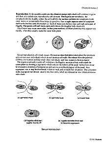
Life Cycle of Chlamydomonas (With Diagram) - Biology Discussion The sexual reproduction in Chlamydomonas can be isogamous, anisogamous or oogamous. he thallus can be homothallic i.e., both types of gametes are produced in same thallus e.g., C. mogama and C. media or can be heterothallic i.e., (+) and (-) gametes come from different parents, he gametes may be naked and called gymnogametes e.g., C. debaryana o...
[Groenwieren: Green algae: Chlorophytae
Chlamydomonas | Facts, Structure, Life Cycle, & Classification Chlamydomonas, genus of biflagellated single-celled green algae (family Chlamydomonadaceae) found in soil, ponds, and ditches. Chlamydomonas species can become so abundant as to colour fresh water green, and one species, C. nivalis, contains a red pigment known as hematochrome, which sometimes imparts a red colour to melting snow.

Chlamydomonas | ClipArt ETC
Use this labeled diagram of a chlamydomonas cell to - Course Hero Use this labeled diagram of a Chlamydomonas cell to address the following two questions. 32. Which of the following statements correctly identifies aspects related to photosynthesis and/or respiration? 1. Acetyl CoA is most often found in G. 2. NADPH accumulates in C. 3. ATP is found in F. 4.

Untitled | Pearltrees
About Chlamydomonas - Chlamydomonas Resource Center Chlamydomonas is a genus of unicellular green algae (Chlorophyta). These algae are found all over the world, in soil, fresh water, oceans, and even in snow on mountaintops. Algae in this genus have a cell wall, a chloroplast, an "eye" that perceives light and two anterior flagella with which they can swim using a breast-stroke type motion.

33 best Protists images on Pinterest | Ap biology, Biology lessons and Life science
Chlamydomonas reinhardtii - an overview | ScienceDirect Topics Chlamydomonas reinhardtii cells are oval shaped, c. 10 μm in length and 3 μm in width, with two flagellae at their anterior end (Figure 1). The cells contain a single chloroplast occupying 40% of the cell volume and several mitochondria. ... Diagram labeling densities in the averaged image. (B) Image average from thin sections of pf14 ...

Labelled Diagram Of Chlamydomonas - Top Label Maker
Draw a neat labelled diagram. Chlamydomonas - Biology Draw a neat labelled diagram. Chlamydomonas . Maharashtra State Board HSC Science (General) 11th. Textbook Solutions 8018. Important Solutions 19. Question Bank Solutions 5546. Concept Notes & Videos 439. Syllabus. Advertisement Remove all ads. Draw a neat labelled diagram. ...
Chlamydomonas | ClipArt ETC
Chlamydomonas Photos and Premium High Res Pictures - Getty Images Diagram of Chlamydomonas angulosa, Flagellated Protozoan. Drawing. Chlamydomonas, illustration. snow alga or snow algae -chlamydomonas nivalis-, kaunergrat range at the back, seeles see lake, kaunertal valley, tyrol, austria - chlamydomonas stock pictures, royalty-free photos & images.
Structure Used By Amoeba For Movement
Chlamydomonas as a Model Organism - Rice University Chlamydomonas as a Model Organism. Chlamydomonas, a genus of unicellular photosynthetic flagellates, is an important model for studies of such fundamental processes as photosynthesis, motility, responses to stimuli such as light, and cell-cell recognition.C. reinhardi, the most commonly studied species of Chlamydomonas, has a relatively simple genome, which has been sequenced.
Post a Comment for "42 chlamydomonas diagram with labels"