41 the brain with labels
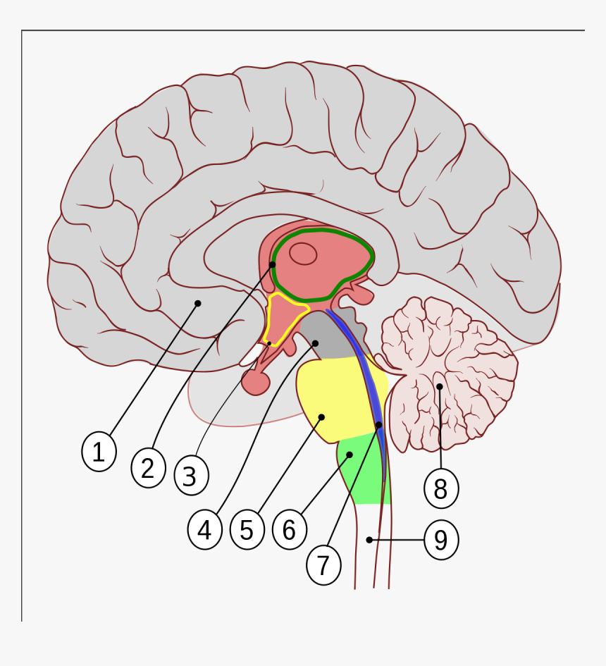
1628 Brain diagram with labels Images, Stock Photos & Vectors 1,628 brain diagram with labels stock photos, vectors, and illustrations are available royalty-free. See brain diagram with labels stock video clips. Brain: Anatomy, Pictures, Functions, and Conditions The brainstem is an area located at the base of the brain that contains structures vital for involuntary functions such as the heartbeat and breathing. The brain stem is comprised of the midbrain, pons, and medulla. 3 Midbrain The midbrain is often considered the smallest region of the brain.
Amazon.com: XINDAM 3D Human Brain with Labels Anatomical Model ... This item: XINDAM 3D Human Brain with Labels Anatomical Model Paperweight (Laser Etched) in Crystal Glass Ball Science Gift (Included LED Base) $66.99 Brain 11 Ounce Ceramic Coffee Mug (WC462M) $16.98 Anatomic Brain Specimen Coasters (Set of 10) - Neuroscience Gifts, Gifts for Medical Student Gifts Brain Decor Human Anatomy Gifts
The brain with labels
Label the Brain Anatomy Diagram Flashcards | Quizlet Start studying Label the Brain Anatomy Diagram. Learn vocabulary, terms, and more with flashcards, games, and other study tools. Nervous System - Label the Brain Nervous System - Label the Brain Nervous System - Brain Name: Choose the correct names for the parts of the brain. ( 1) (2) (3) (4) (5) (6) (7) (8) ( 9) This brain part controls thinking. (10) This brain part controls balance, movement, and coordination. (11) This brain part controls involuntary actions such as breathing, heartbeats, and digestion. DOC Label the Brain Anatomy Diagram Answers: Label the Brain Diagram The Brain. Read the definitions below, then label the brain anatomy diagram. Cerebellum - the part of the brain below the back of the cerebrum. It regulates balance, posture, movement, and muscle coordination. Corpus Callosum - a large bundle of nerve fibers that connect the left and right cerebral hemispheres.
The brain with labels. 2,791 Labeled brain anatomy Images, Stock Photos & Vectors - Shutterstock 2,791 labeled brain anatomy stock photos, vectors, and illustrations are available royalty-free. See labeled brain anatomy stock video clips Image type Orientation Artists Sort by Popular Healthcare and Medical Anatomy Icons and Graphics human brain brain organ medicine cerebral cortex cerebellum limbic system Next of 28 101 Labeled Brain Images and a Consistent Human Cortical ... We introduce the Mindboggle-101 dataset, the largest and most complete set of free, publicly accessible, manually labeled human brain images. To manually label the macroscopic anatomy in magnetic resonance images of 101 healthy participants, we created a new cortical labeling protocol that relies on robust anatomical landmarks and minimal manual edits after initialization with automated labels ... Labeled Brain Model Diagram | Science Trends The cerebrum is the largest and most complex portion of the human brain. The cerebrum's function is to control our actions and thoughts, either conscious or unconscious, and responses to stimuli. The cerebrum itself is typically divided into four different lobes: the temporal lobe, the parietal lobe, the occipital lobe, and the frontal lobe. Positions and Functions of the Four Brain Lobes - MD-Health.com The brain is divided into four sections, known as lobes (as shown in the image). The frontal lobe, occipital lobe, parietal lobe, and temporal lobe have different locations and functions that support the responses and actions of the human body. Let's start by identifying where each lobe is positioned in the brain. Position of the Lobes
28923 Brain labels Images, Stock Photos & Vectors Find Brain labels stock images in HD and millions of other royalty-free stock photos, illustrations and vectors in the Shutterstock collection. Brain Anatomy and How the Brain Works - Hopkins Medicine The cerebellum ("little brain") is a fist-sized portion of the brain located at the back of the head, below the temporal and occipital lobes and above the brainstem. Like the cerebral cortex, it has two hemispheres. The outer portion contains neurons, and the inner area communicates with the cerebral cortex. Label The Brain - Mr. Barth's Class You won't label the parts of the brain on this website, but you'll familiarize yourself with the location of the parts and their basic functions. Lobes of the Brain Click on the link to the left to label the lobes of the brain. See how quickly you can do it with 100% accuracy. Lobes and Neuron Diagram The Human Brain - Visible Body The brain gives us self-awareness and the ability to speak and move in the world. Its four major regions make this possible: The cerebrum, with its cerebral cortex, gives us conscious control of our actions. The diencephalon mediates sensations, manages emotions, and commands whole internal systems. The cerebellum adjusts body movements, speech ...
Brain Label - The Biology Corner Image of the brain showing its major features for students to practice labeling. Answers are included. Related 3D Models for 3d model the brain with labels ID: 700274 , 1,984 views , Tags: 3d models, anatomy, brain, head, human, label, medical Description: This is a 3d model of the brain with labels,which tells us the information of the brain.This model is sliced into parts frontal lobe, motor cortex, etc. Label The Brain Worksheets & Teaching Resources | TpT The Primary Brain 170 $3.25 Digital Download PPTX (2.65 MB) Organize your classroom library bins with accelerated reader labels. These black and brights AR labels are EDITABLE so you can customize your book bin labels for a classroom library. Just open the PowerPoint and add in your own library label text. Brain Label (Remote) - The Biology Corner The activity includes an external view of the brain where students label the lobes of the cerebrum (frontal, parietal, occipital, and temporal) and the cerebellum. Next students drag and drop labels to the internal structures, such as the thalamus, midbrain, corpus callosum, pineal body, and colliculi.
30 Label Of The Brain - Labels Information List
The Brain - Diagram and Explanation The outer 3 millimeters of "gray matter" is the cerebral cortex which consists of closely packed neurons that control most of our body functions, including the mysterious state of consciousness, the senses, the body's motor skills, reasoning and language.

Label the Brain Quiz
Label Brain Diagram Printout - EnchantedLearning.com The Brain. Read the definitions below, then label the brain anatomy diagram. Cerebellum - the part of the brain below the back of the cerebrum. It regulates balance, posture, movement, and muscle coordination. Corpus Callosum - a large bundle of nerve fibers that connect the left and right cerebral hemispheres.
Brain Viewed from Above | ClipArt ETC
Parts of the brain: Learn with diagrams and quizzes | Kenhub Labeled brain diagram First up, have a look at the labeled brain structures on the image below. Try to memorize the name and location of each structure, then proceed to test yourself with the blank brain diagram provided below. Labeled diagram showing the main parts of the brain Blank brain diagram (free download!)

Brain WebQuest - GW8science
Brain Records - Wikipedia Brain was a Hamburg-based record label prominent in the 1970s releasing several important Krautrock records by bands such as Neu!, Cluster and Guru Guru.Many of its more prominent records are currently being reissued on CD by Repertoire Records.. In the middle of 1971, Rolf-Ulrich Kaiser's management style at Ohr caused two of his A&R men, Bruno Wendel and Günter Körber, to leave Ohr and set ...

Pin on Embellish(-ing the truth)
Brain (Human Anatomy): Picture, Function, Parts, Conditions, and More The brain is also divided into several lobes: • The frontal lobes are responsible for problem solving and judgment and motor function. • The parietal lobes manage sensation, handwriting, and body...

Name
Labeled Diagrams of the Human Brain You'll Want to Copy Now Labeled Diagrams of the Human Brain Central Core The central core consists of the thalamus, pons, cerebellum, reticular formation and medulla. These five regions are the central areas that regulate breathing, pulse, arousal, balance, sleep and early stages of processing sensory information.

The Brain | Teaching Resources
Amazon.com: brain model labeled VEVOR Human Brain Model Anatomy 4-Part Model of Brain w/Labels & Display Base Color-Coded Life Size Human Brain Anatomical Model Brain Teaching Human Brain for Science Classroom Study Display Model. 3.4 out of 5 stars 3. $159.19 $ 159. 19. Get it Wed, Mar 30 - Mon, Apr 4. FREE Shipping.

Animals And Birds: Badger animal profile 2011 & Pic`s
3D Brain This interactive brain model is powered by the Wellcome Trust and developed by Matt Wimsatt and Jack Simpson; reviewed by John Morrison, Patrick Hof, and Edward Lein. Structure descriptions were written by Levi Gadye and Alexis Wnuk and Jane Roskams .
Online Exhibits - Crafting Persuasion
Label Parts of the Brain Quiz - PurposeGames.com An unregistered player played the game 4 hours ago About this Quiz This is an online quiz called Label Parts of the Brain There is a printable worksheet available for download here so you can take the quiz with pen and paper. From the quiz author Okay. This quiz has tags. Click on the tags below to find other quizzes on the same subject. biology
Immensely Satisfying Animated Gifs by Nicolas Fong – BOOOOOOOM! – CREATE * INSPIRE * COMMUNITY ...
Labels on the Brain - Cognitioneducation We love labels for what they do well — they make things easy for us. Labels are a product of the way our minds work - in fact, this process may be one of our brains greatest feats. Our brain circuitry pattern-matches like nobody's business; doing so affords a necessary level of simplicity in an otherwise overly complex world.
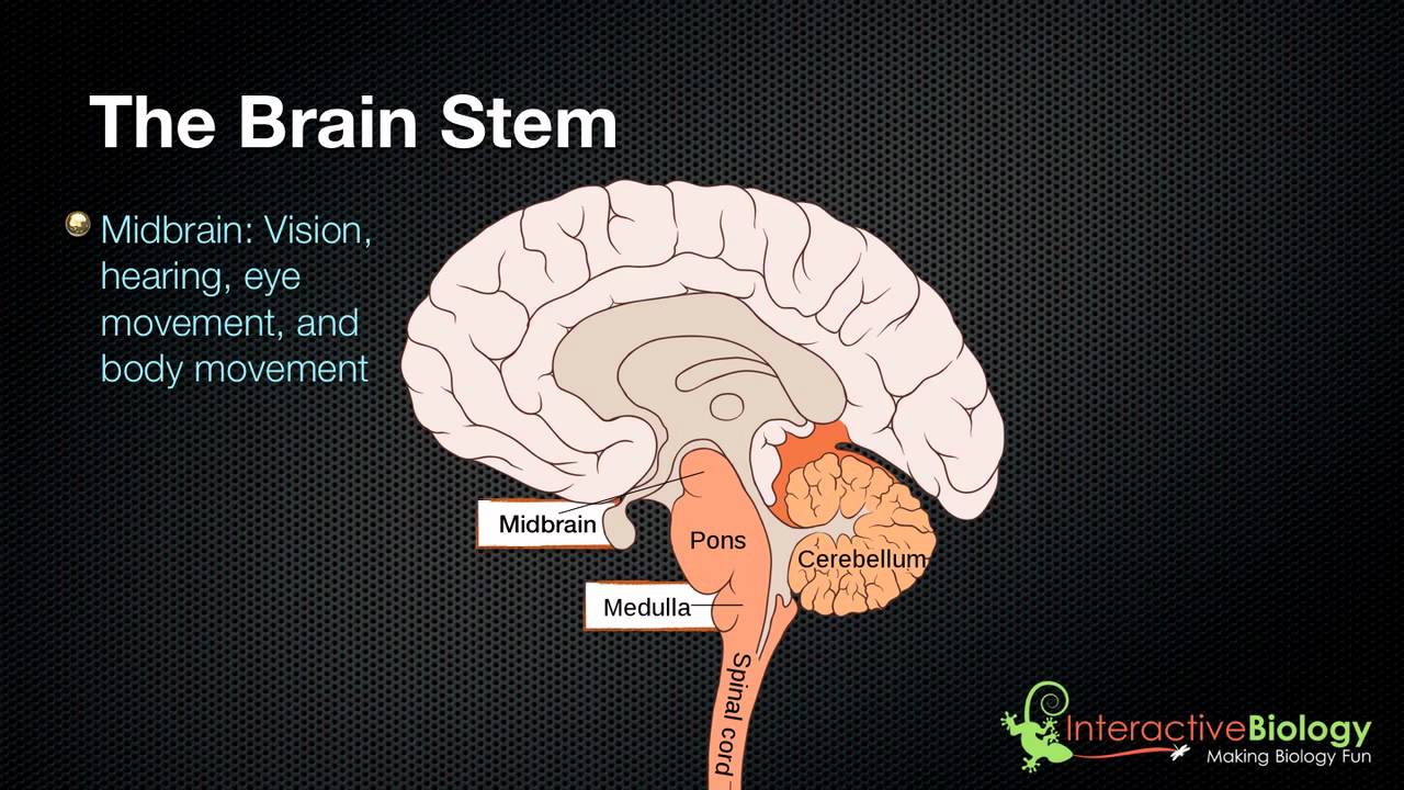
027 The 3 parts of the brain stem and their functions - YouTube
Intracerebroventricular infusion of CHO5, a rat monoclonal antibody ... 1 Paul Flechsig Institute for Brain Research, Department of Neurochemistry, University of Leipzig, Germany. rossn@medizin.uni-leipzig.de; PMID: ... Using double labeling immunocytochemistry, the rat monoclonal antibody CHO5 against mouse p75NTR was found to label mouse basal forebrain neurons, which also demonstrated immunoreactivity for ...

Building the Brain for Literacy with SPELL-Links - Learning By Design
28926 Brain label Images, Stock Photos & Vectors - Shutterstock Find Brain label stock images in HD and millions of other royalty-free stock photos, illustrations and vectors in the Shutterstock collection.
Laboratory Charts and Posters: Brain
Lobes of the brain: Structure and function | Kenhub The brain, along with the spinal cord, is the main organ of the central nervous system. It is the most complex organ of the body, with many layers and components that play their roles in almost every function performed by the body. The brain is composed of the cerebrum, cerebellum and brainstem.

Lateral View of Optic Radiation of Left Hemisphere | Neuroanatomy | The Neurosurgical Atlas, by ...
709 results for labeled brain in images - Adobe Stock Search from thousands of royalty-free Labeled Brain stock images and video for your next project. Download royalty-free stock photos, vectors, ...

33 Human Brain With Label - Labels Database 2020
Brain Label | Human anatomy and physiology, Basic anatomy and ... Brain Label Image of the brain showing its major features for students to practice labeling. Answers are included. Biologycorner 17k followers More information Brain Label Find this Pin and more on Anatomy & Physiology by Page Johnson. Basic Anatomy And Physiology Brain Anatomy Science Education Physical Science Science Experiments

Mr. Forde - Life Science
DOC Label the Brain Anatomy Diagram Answers: Label the Brain Diagram The Brain. Read the definitions below, then label the brain anatomy diagram. Cerebellum - the part of the brain below the back of the cerebrum. It regulates balance, posture, movement, and muscle coordination. Corpus Callosum - a large bundle of nerve fibers that connect the left and right cerebral hemispheres.

brain-anatomy - The Mind Voyager
Nervous System - Label the Brain Nervous System - Label the Brain Nervous System - Brain Name: Choose the correct names for the parts of the brain. ( 1) (2) (3) (4) (5) (6) (7) (8) ( 9) This brain part controls thinking. (10) This brain part controls balance, movement, and coordination. (11) This brain part controls involuntary actions such as breathing, heartbeats, and digestion.

How many Animals can you see | Whatsapp Puzzles world, Quiz, Games, Riddles and messages
Label the Brain Anatomy Diagram Flashcards | Quizlet Start studying Label the Brain Anatomy Diagram. Learn vocabulary, terms, and more with flashcards, games, and other study tools.
Post a Comment for "41 the brain with labels"