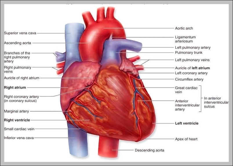
42 structure of the heart with labels
› metal-hammerMetal Hammer | Louder 1 day ago · Subscribe to the world's greatest music magazines; Try a single issue or save on a subscription; Issues delivered straight to your door or device Structure of the Heart | SEER Training The human heart is a four-chambered muscular organ, shaped and sized roughly like a man's closed fist with two-thirds of the mass to the left of midline. The heart is enclosed in a pericardial sac that is lined with the parietal layers of a serous membrane. The visceral layer of the serous membrane forms the epicardium. Layers of the Heart Wall
Human Heart Diagram Labeled | Science Trends The human heart usually weighs somewhere between 10 to 12 ounces in men and between 8 to 10 ounces in women, and in terms of size is roughly the size of the fist. The heart has four different chambers: the left and right ventricles and the left and right atriums.

Structure of the heart with labels
Heart Anatomy Labeling Game - PurposeGames.com This is an online quiz called Heart Anatomy Labeling Game There is a printable worksheet available for download here so you can take the quiz with pen and paper. Your Skills & Rank Total Points 0 Get started! Today's Rank -- 0 Today 's Points One of us! Game Points 19 You need to get 100% to score the 19 points available Actions Heart Anatomy: Labeled Diagram, Structures, Function, and Blood Flow Chambers of the Heart Let's begin with the chambers of the heart. There are 4 chambers, labeled 1-4 on the diagram below. To help simplify things, we can convert the heart into a square. We will then divide that square into 4 different boxes which will represent the 4 chambers of the heart. Human Heart - Diagram and Anatomy of the Heart - Innerbody The heart is a muscular organ about the size of a closed fist that functions as the body's circulatory pump. It takes in deoxygenated blood through the veins and delivers it to the lungs for oxygenation before pumping it into the various arteries (which provide oxygen and nutrients to body tissues by transporting the blood throughout the body).
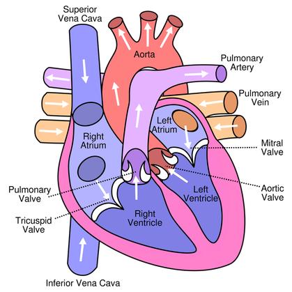
Structure of the heart with labels. Human Heart (Anatomy): Diagram, Function, Chambers, Location in Body Human Heart (Anatomy): Diagram, Function, Chambers, Location in Body The right atrium receives blood from the veins and pumps it to the right ventricle. The right ventricle receives blood from the... Label the heart — Science Learning Hub Label the heart Interactive Add to collection In this interactive, you can label parts of the human heart. Drag and drop the text labels onto the boxes next to the diagram. Selecting or hovering over a box will highlight each area in the diagram. Right ventricle Right atrium Left atrium Pulmonary artery Left ventricle Pulmonary vein Semilunar valve The Anatomy of the Heart, Its Structures, and Functions The heart is the organ that helps supply blood and oxygen to all parts of the body. It is divided by a partition (or septum) into two halves. The halves are, in turn, divided into four chambers. The heart is situated within the chest cavity and surrounded by a fluid-filled sac called the pericardium. This amazing muscle produces electrical ... Structure of the Heart | The Franklin Institute Structure of the Heart Although most people know that the human heart doesn't bear much resemblance to a heart drawn on a Valentine's Day card, the image can still be a useful way to learn and remember the parts of the heart. The heart consists of four chambers: two atria on the top and two ventricles on the bottom.
Label Internal Anatomy of The Heart Diagram | Quizlet Start studying Label Internal Anatomy of The Heart. Learn vocabulary, terms, and more with flashcards, games, and other study tools. A Labeled Diagram of the Human Heart You Really Need to See The human heart, comprises four chambers: right atrium, left atrium, right ventricle and left ventricle. The two upper chambers are called the left and the right atria, and the two lower chambers are known as the left and the right ventricles. The two atria and ventricles are separated from each other by a muscle wall called 'septum'. The Anatomy of the Heart - Quiz 1 - Free Anatomy Quiz The circulatory system - lower body image, with blank labels attached. The circulatory system - a PDF file of the upper and lower body for printing out to use off-line. Describe and explain the function of the circulatory system - The circulatory system consists of the heart, the blood vessels (veins, arteries, and capillaries), and the blood. › regulatory-information › search-fdaSmall Entity Compliance Guide on Structure/Function Claims Finally, the preamble to this rule clarifies several legal issues that are important to understand if you use structure/function claims on the labels or in the labeling of your products. They are ...
Heart Blood Flow | Simple Anatomy Diagram, Cardiac Circulation ... - EZmed Step 1 and 6 involve a blood vessel, which makes sense as this is how blood enters and exits that side of the heart. Steps 2-5 involve a chamber, valve, chamber, and valve. So if you remember this general pattern, it will help you recall the order in which blood flows through each side of the heart. Heart anatomy: Structure, valves, coronary vessels | Kenhub The heart is shaped as a quadrangular pyramid, and orientated as if the pyramid has fallen onto one of its sides so that its base faces the posterior thoracic wall, and its apex is pointed toward the anterior thoracic wall. Heart Diagram with Labels and Detailed Explanation - BYJUS Diagram of Heart. The human heart is the most crucial organ of the human body. It pumps blood from the heart to different parts of the body and back to the heart. The most common heart attack symptoms or warning signs are chest pain, breathlessness, nausea, sweating etc. The diagram of heart is beneficial for Class 10 and 12 and is frequently ... How to Draw the Internal Structure of the Heart (with Pictures) 1. To find a good diagram, go to Google Images, and type in "The Internal Structure of the Human Heart". Find an image that displays the entire heart, and click on it to enlarge it. 2. Find a piece of paper and something to draw with. Start with the pulmonary veins.
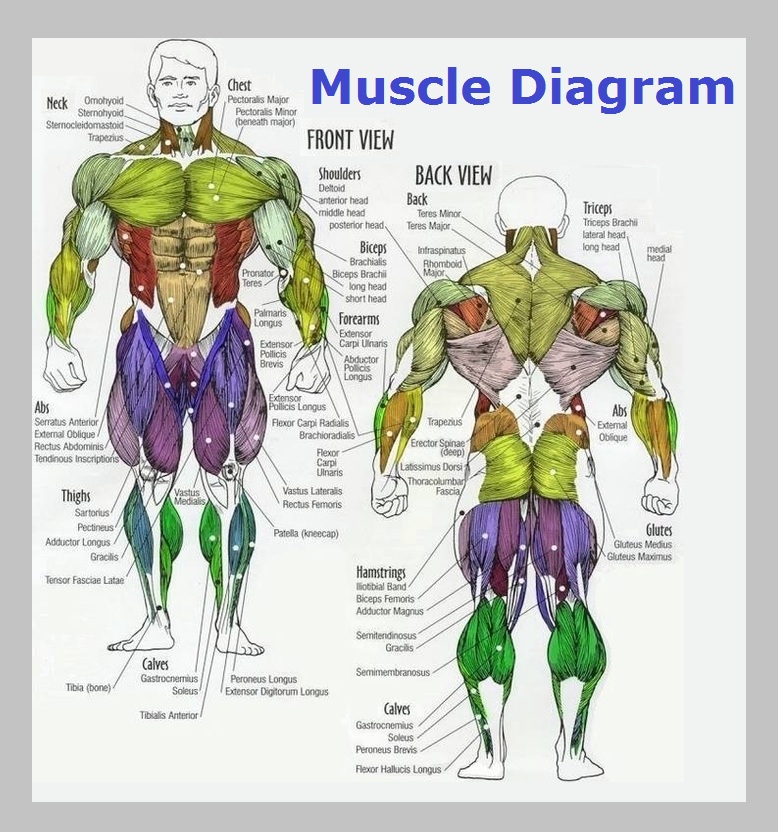
Muscle Diagram – Graph Diagram
› site-mapAutoblog Sitemap Here's how to disable adblocking on our site. Click on the icon for your Adblocker in your browser. A drop down menu will appear. Select the option to run ads for autoblog.com, by clicking either ...
12+ Model Heart Labeled | Robhosking Diagram
Labelling the heart — Science Learning Hub Labelling the heart — Science Learning Hub Labelling the heart Add to collection The heart is a muscular organ that pumps blood through the blood vessels of the circulatory system. Blood transports oxygen and nutrients to the body. It is also involved in the removal of metabolic wastes. Topics Concepts Citizen science Teacher PLD Glossary Sign in
heart - a level biology student
| The Heart Foundation The Heart Foundation saves lives and improves health through funding world-class cardiovascular research, guidelines for health professionals, informing the public and assisting people with cardiovascular disease
Congenital Heart Defects - How the Heart Works | CDC
Label Heart Anatomy Diagram Printout - EnchantedLearning.com Oxygen-poor blood enters the right atrium of the heart (via veins called the inferior vena cava and the superior vena cava). The blood is then pumped into the right ventricle and then through the pulmonary artery to the lungs, where the blood is enriched with oxygen (and loses carbon dioxide).

Cuthbert - 7th Grade Science Day to Day: Comparing Plant and Animal Cells
147 Heart Anatomy With Labels Premium High Res Photos Browse 147 heart anatomy with labels stock photos and images available, or start a new search to explore more stock photos and images. of 3. NEXT.
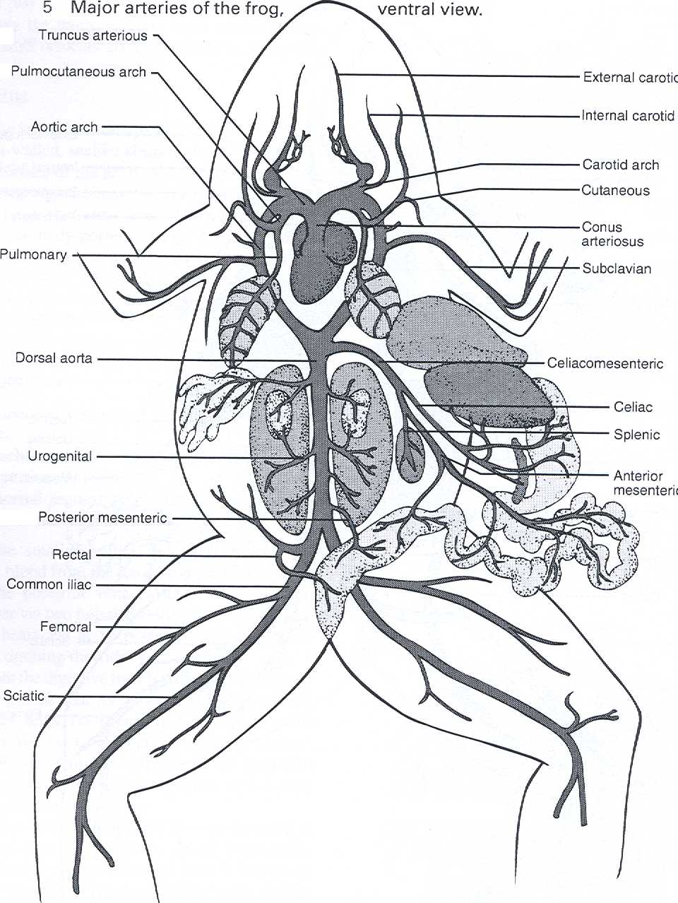
Dissection of the Frog
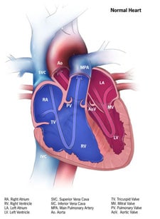
Heart Diagram with Labels and Detailed Explanation The heart is located under the ribcage, between the lungs and above the diaphragm. It weighs about 10.5 ounces and is cone shaped in structure. It consists of the following parts: Heart Detailed Diagram Heart - Chambers There are four chambers of the heart . The upper two chambers are the auricles and the lower two are called ventricles.
Heart Structure and Function
Ch. 19 Circulatory System- heart Flashcards | Quizlet Place the labels in order denoting the flow of blood through the pulmonary circuit beginning with the right atrium and ending in the left atrioventricular valve. The first and last structures are given. Right atrium 1. tricuspid valve 2. right ventricle 3. pulmonary valve 4. pulmonary trunk 5. pulmonary artery 6. lungs 7. pulmonary vein

Labeling the Heart
ods.od.nih.gov › factsheets › Calcium-ConsumerCalcium - Consumer Almost all calcium in the body is stored in bones and teeth, giving them structure and hardness. Your body needs calcium for muscles to move and for nerves to carry messages between your brain and every part of your body. Calcium also helps blood vessels move blood throughout your body and helps release hormones that affect many functions in ...

Anatomy and Physiology 2 Eportfolio: Objective 24: Structures of the Heart
uxplanet.org › designing-more-efficient-formsDesigning More Efficient Forms: Structure, Inputs, Labels and ... Apr 10, 2016 · Labels. Clear label text is one of the primary ways to make UIs more accessible. Labels tell the user the purpose of the field and they should remain visbile even after completing the field. Number of Words. Labels are not help texts. You should use succinct, short and descriptive labels (a word or two) so users can quickly scan your form.
32 Label The Diagram Of The Heart - Label Design Ideas 2020
Diagrams, quizzes and worksheets of the heart | Kenhub Worksheet showing unlabelled heart diagrams. Using our unlabeled heart diagrams, you can challenge yourself to identify the individual parts of the heart as indicated by the arrows and fill-in-the-blank spaces. This exercise will help you to identify your weak spots, so you'll know which heart structures you need to spend more time studying ...

Label Heart Structure | Medical Science Navigator
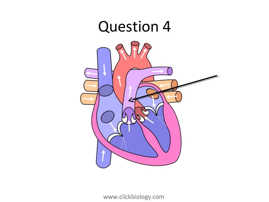
Heart Labeling Quiz: How Much You Know About Heart Labeling? Here is a Heart labeling quiz for you. The human heart is a vital organ for every human. The more healthy your heart is, the longer the chances you have of surviving, so you better take care of it. Take the following quiz to know how much you know about your heart. Questions and Answers 1. What is #1? 2. What is #2? 3. What is #3? 4. What is #4?

The Heart | S-cool, the revision website
What part of the heart is Labelled? - Ufoscience.org Label the heart. In this interactive, you can label parts of the human heart. Drag and drop the text labels onto the boxes next to the diagram. Selecting or hovering over a box will highlight each area in the diagram. In this interactive, you can label parts of the human heart. Drag and drop the text labels onto the boxes next to the heart diagram.

Anterior View of Sheep Heart and Structures | Anatomy organs, Cardiology, Medical
A Diagram of the Heart and Its Functioning Explained in Detail The heart blood flow diagram (flowchart) given below will help you to understand the pathway of blood through the heart.Initial five points denotes impure or deoxygenated blood and the last five points denotes pure or oxygenated blood. 1.Different Parts of the Body. ↓. 2.Major Veins.
The Anatomy and Physiology of Animals/Circulatory System Worksheet - WikiEducator
webaim.org › standards › wcagWebAIM's WCAG 2 Checklist Feb 26, 2021 · 2.4.6 Headings and Labels (Level AA) Page headings and labels for form and interactive controls are informative. Avoid duplicating heading (e.g., "More Details") or label text (e.g., "First Name") unless the structure provides adequate differentiation between them. 2.4.7 Focus Visible (Level AA)

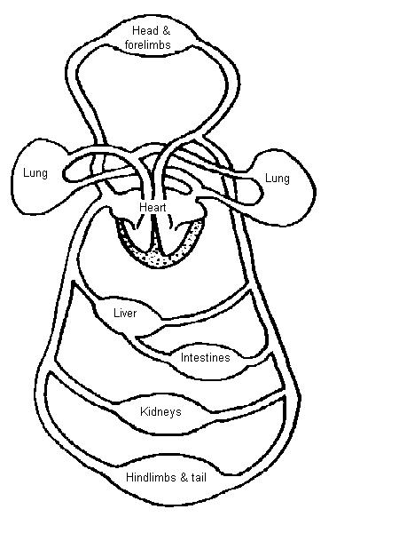
Circulatory function
The Human Heart Without Labels at Anatomy Close up black and white heart anatomy with labels graphic design. Source: clipart-library.com. Flaps that prevent backflow of blood. On average it beats about 100,000 times a day and pumps 7,571 liters (2,000 gallons) of blood. The human heart is the pump for the circulatory system, and along with the circulatory system is considered to be an ...
35 Label The Diagram Of The Heart - Labels Design Ideas 2020
Label the Heart - The Biology Corner Shows a picture of a heart with letters and blanks for practice with labeling the parts of the heart and tracing the flow of blood within the heart.

In this diagram they are showing the function of the heart as they have labels to the ...
Diagram of Human Heart and Blood Circulation in It Exterior of the Human Heart A heart diagram labeled will provide plenty of information about the structure of your heart, including the wall of your heart. The wall of the heart has three different layers, such as the Myocardium, the Epicardium, and the Endocardium. Here's more about these three layers. Epicardium
Post a Comment for "42 structure of the heart with labels"