38 cell membrane diagram with labels
Animal cells and plant cells - Cells to systems - BBC Bitesize Part Function Found in; Cell membrane: Controls the movement of substances into and out of the cell: Plant and animal cells: Cytoplasm: Jelly-like substance, where chemical reactions happen Cell Membrane Diagram Labeled : Functions and Diagram The plasma membrane is a protective barrier that surrounds the interior of the cell. We are pleased to provide you with the picture named Cell Membrane Diagram. Diploma Biology - Core, Topic 2, 2.4 Cell Membranes …. PPT - The Cell Membrane PowerPoint Presentation - ID:501873.
Basic Cell Membrane Label - Labelled diagram - Wordwall Integral Protein (channel), Peripheral Protein, Phosphate, Lipid, Hydrophilic, Hydrophobic, Glycoprotein.

Cell membrane diagram with labels
Cell Membrane Is Labeled : Functions and Diagram The cell membrane All cells are enclosed by a cell membrane. The cell membrane is a thin flexible layer around the cells of all living things. One of the foremost intricate duties that healthiness authorities face throughout their interplay with patients is helping them realise the problems and a way to motivate them in regards to the diagnosis and treatment available. All of this has been made much less complicated because of the assistance of human anatomy diagrams. Cell Membrane Is Diagram of a cell membrane with labels | NIST Biology in Reflectometry. Essential Biological Functions. Immune response, Cell metabolism, Neurotransmission, Photosynthesis, Cell adherence, Cell growth and differentiation. Potential Commercial Applications. Drug response monitoring, Chemical manufacturing, Biosensing, Energy conversion, Tissue engineering. Cell Membrane Labeled Simple : Functions and Diagram Bacteria diagram additionally indicates Periplasmic space, that is a cellular compartment found in simple terms in bacteria that have an outer membrane and a plasma membrane . 35 Label The Plasma Membrane - Labels Database 2020 (Chris Cooper) Similarly, nothing can enter the cell. It is the outermost part of the cell in animals.
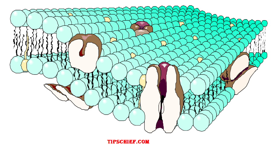
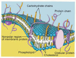
Cell membrane diagram with labels. PDF Membrane Structure and Function - Phoenix College Major Components of the Cell Membrane: Lipids • Phospholipids are amphipathicmolecules (with hydrophobictails and a hydrophilichead) • One of the phospholipid tails exist mostly in a transconfiguration, providing more fluidityto the membrane • Cholesterol is a rigid molecule that makes membranes less fluid Cholesterol PDF Human Cell Diagram, Parts, Pictures, Structure and Functions 2 Diagram of the human cell illustrating the different parts of the cell. Cell Membrane The cell membrane is the outer coating of the cell and contains the cytoplasm, substances within it and the organelle. It is a double-layered membrane composed of proteins and lipids. Cell Membrane Made Up Of Labeled : Functions and Diagram Cell Membrane Made Up Of Labeled Tuesday, February 2nd 2021. | Diagram Cell Membrane Made Up Of. Their proportions vary between different types of eukaryotic cells, but their basic characteristics remain the same. Phospholipids are lipid molecules made up of a phosphate group head and two fatty acid tails. Label Cell Membrane Diagram | Quizlet Start studying Label Cell Membrane. Learn vocabulary, terms, and more with flashcards, games, and other study tools.
PDF Cell Membrane Structure (1.3) - University of São Paulo Cell Membrane Structure (1.3) IB Diploma Biology Essential idea: The structure of biological ... When drawing a diagram of a phospholipid this is a good example which shows all the key features 1.3.1 Phospholipids form bilayers in water due to the amphipathic ... • Labels clearly written • (Scale bar if appropriate) ... CELL MEMBRANE LABEL Diagram | Quizlet Practice labeling the parts of the cell membrane Terms in this set (6) Channel Protein hole or tunnel that particles may pass through to go in / out of cell Marker protein identifies or labels the cell Receptor protein receives information Heads part of the phospholipid that loves water (hydrophili) - points to the most outside and inside of cell History of cell membrane theory - Wikipedia Two experiments in 1924 laid the groundwork to fill in this gap. By measuring the capacitance of erythrocyte solutions Fricke determined that the cell membrane was 3.3 nm thick. Although the results of this experiment were accurate, Fricke misinterpreted the data to mean that the cell membrane is a single molecular layer. Animal Cells: Labelled Diagram, Definitions, and Structure The endoplasmic reticulum (s) are organelles that create a network of membranes that transport substances around the cell. They have phospholipid bilayers. There are two types of ER: the rough ER, and the smooth ER. The rough endoplasmic reticulum is rough because it has ribosomes (which is explained below) attached to it.
A Well-labelled Diagram Of Animal Cell With Explanation Diagram Of Animal Cell. Animal cells are eukaryotic cells that contain a membrane-bound nucleus. They are different from plant cells in that they do contain cell walls and chloroplast. The animal cell diagram is widely asked in Class 10 and 12 examinations and is beneficial to understand the structure and functions of an animal. Cell Bio - Ch. 22 Flashcards | Quizlet The diagram below shows the five main transport proteins that control the distribution of Na+ and K+ ions across the plasma membrane of an axon. Assume that the membrane is at resting potential---the membrane potential of the axon remains constant at about -70 mV. Structure of Membrane in Cells (With Diagram) - Biology Discussion Cell walls are formed through apposition, i.e., the wall material is deposited by the protoplast on the plasma membrane. The first cell wall (Primary wall) is formed during the cell growth phase. When the cell elongation is stopped the secondary cell wall, formation starts (Fig. 2.13). Cell Organelles- Definition, Structure, Functions, Diagram A cell wall is multilayered with a middle lamina, a primary cell wall, and a secondary cell wall. The middle lamina contains polysaccharides that provide adhesion and allow binding of the cells to one another. After the middle lamina is the primary cell wall which is composed of cellulose.

Bacteria Diagram
Animal Cell Diagram with Label and Explanation: Cell ... - Collegedunia The cell membrane is the thin semi-permeable layer surrounding the cell; its main functionality is to protect the cell. It has hair-like structures cilia and flagella on it. The cell membrane controls the entry and exit of nutrients into the animal cell. Cytoplasm

Simple Cuboidal
Cell Membrane With Labels Labeled : Functions and Diagram Cell Membrane With Labels. It protects the integrity of the cell along with supporting the cell and helping to maintain the cell's shape. A cell wall is multilayered with a middle lamina, a primary cell wall, and a. We all remember that the human physique is very intricate and a technique I learned to are aware of it is via the manner of human ...

Root Hair Cell - Biology
Labeled Plant Cell With Diagrams - Science Trends The parts of a plant cell include the cell wall, the cell membrane, the cytoskeleton or cytoplasm, the nucleus, the Golgi body, the mitochondria, the peroxisome's, the vacuoles, ribosomes, and the endoplasmic reticulum. Parts Of A Plant Cell The Cell Wall Let's start from the outside and work our way inwards.

31 Cell Membrane Diagram To Label - Labels For You
Plant Cells: Labelled Diagram, Definitions, and Structure Plants have a rigid cell wall that surrounds the plasma membrane. The cell wall is made of cellulose and lignin, which are strong and tough compounds. Plant Cells Labelled Plastids and Chloroplasts Plants make their own food through photosynthesis. Plant cells have plastids, which animal cells don't.
Biology diagram worksheet
The Cell - ScienceQuiz.net The diagram shows a plant cell as seen under a microscope. Two of the labels are incorrect. What are they? ... A is the cell membrane and DNA is located inside B.?

Cell Membrane Facts Labeled - Cell Diagram
Labeling a cell membrane Diagram | Quizlet Start studying Labeling a cell membrane. Learn vocabulary, terms, and more with flashcards, games, and other study tools.

Biology Diagram Show Structure Of Cell Membrane Stock Illustration - Download Image Now - iStock
Wikipedia:Featured picture candidates/Cell membrane ... Cell membrane (diagrammatic) Original - The cell membrane, also called the plasma membrane or plasmalemma, is a semipermeable lipid bilayer common to all living cells. It contains a variety of biological molecules, primarily proteins and lipids, which are involved in a vast array of cellular processes.

Cell Membrane Structure Diagram | Cell Membrane | Biology | Pinterest | See more best ideas ...
Cell membrane with labeled educational structure scheme vector ... 2. Editable Vector .EPS-10 file. 3. High-resolution JPG image. Use for everything except reselling item itself. Description: Cell membrane with labeled educational structure scheme vector illustration. Anatomical closeup drawing with cross section element. Carbohydrate, globular protein or cholesterol location visualization.

Label The Cell Membrane
Labeled Diagram Of Cell Membrane : Prokaryotic Cell Structure Diagram ... The outer covering of the body cells, which maintains homeostatic condition between inside and outside of the cell is called cell membrane. Schematic diagram of a cell membrane membrane structure, cell structure,. It is made up of . Membrane proteins labeled vector illustration. Learn how to find cell towers near you.
Detailed Diagram Models Cell Membrane Stock Vector (Royalty Free) 376416385 - Shutterstock
Interactive Cell Cycle - CELLS alive INTERPHASE. Gap 0. Gap 1. S Phase. Gap 2. MITOSIS . ^ Cell Cycle Overview Cell Cycle Mitosis > Meiosis > Get the Cell Division PowerPoints
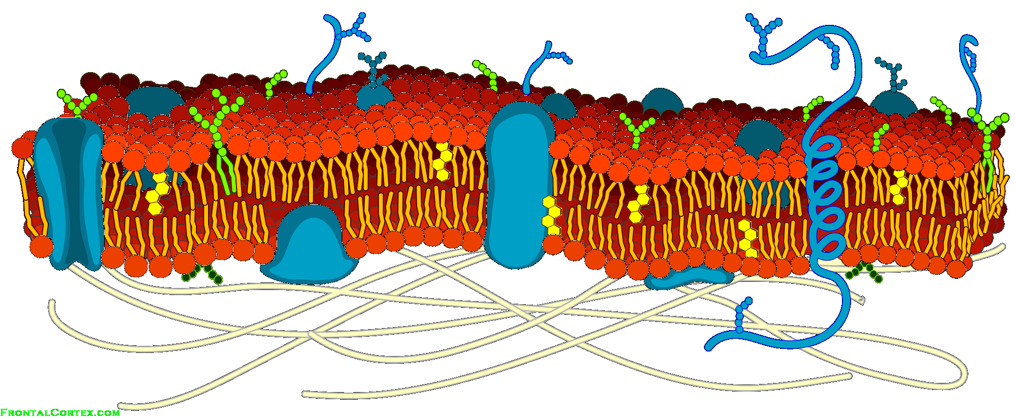
Image: Cell Membrane Detailed Diagram B
03 Label the Cell Diagram | Quizlet Start studying 03 Label the Cell. Learn vocabulary, terms, and more with flashcards, games, and other study tools.
Science KS3 Revision | Flashcards
Label the cell membrane Diagram | Quizlet Start studying Label the cell membrane. Learn vocabulary, terms, and more with flashcards, games, and other study tools.

Animal Cell Labeling
Cell Membrane Vs Cell Wall Labeled : Functions and Diagram Cell membrane helps to enclose the cell organelles and cytosol inside a cell. Cell membrane also helps ions to transfer from the inside to the outside of the cell and vice versa. The cell membrane is also known as the plasma membrane or plasma lemma. It's characterized by a hydrophilic head (glycerol head) and a hydrophobic region that make up the.

Animal Cells Diagram with Labels Awesome Animal Cell Diagrams Labeled | Animal cell project ...
Plasma Membrane Function, Structure & Diagram - Study.com The plasma membrane name comes from its ability to move with the cell in a flexible way, like plasma flowing, and its function in creating a barrier, or membrane to separate the cell from the ...

Explain the nucleus of a cell with a neat labeled diagram - Science - Cell - Structure and ...
Cell Membrane Structure Labeled Labeled : Functions and Diagram Cell Membrane Structure Labeled. Let us study the detailed composition of this lipid bilayer and other substances found in the cell membrane. This structure has two layers, and is represented in the diagram below. We all do not forget that the human physique is very intricate and one way I learned to comprehend it is by way of the style of human ...

Schematic Diagram of a Cell Membrane | Cell Structure Function | Pinterest | Cell membrane, A ...
How to Create 3D Plant Cell and Animal Cell Models for ... Sep 10, 2011 · Organelle: Any specialized structure inside the cell. Cell Membrane: Composed of a double lipid bilayer, the cell membrane separates and protects the cell from its environment, regulates the movement of molecules in and out of the cell, and provides structure to the cell. Cytoplasm: The semifluid substance that fills the cell. All of the cell's ...
Post a Comment for "38 cell membrane diagram with labels"