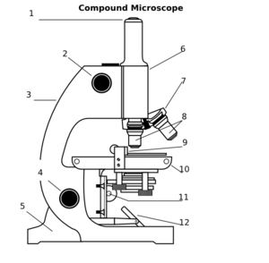
40 compound microscope diagram without labels
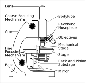
Label Microscope Diagram - EnchantedLearning.com Using the terms listed below, label the microscope diagram. arm - this attaches the eyepiece and body tube to the base. base - this supports the microscope. body tube - the tube that supports the eyepiece. coarse focus adjustment - a knob that makes large adjustments to the focus. diaphragm - an adjustable opening under the stage, allowing ... Compound Microscope- Definition, Labeled Diagram, Principle, Parts, Uses Alternatively, the magnification of the compound microscope is given by: m = D/ fo * L/fe where, D = Least distance of distinct vision (25 cm) L = Length of the microscope tube fo = Focal length of the objective lens fe = Focal length of the eye-piece lens Parts of a Compound Microscope Eyepiece And Body Tube.
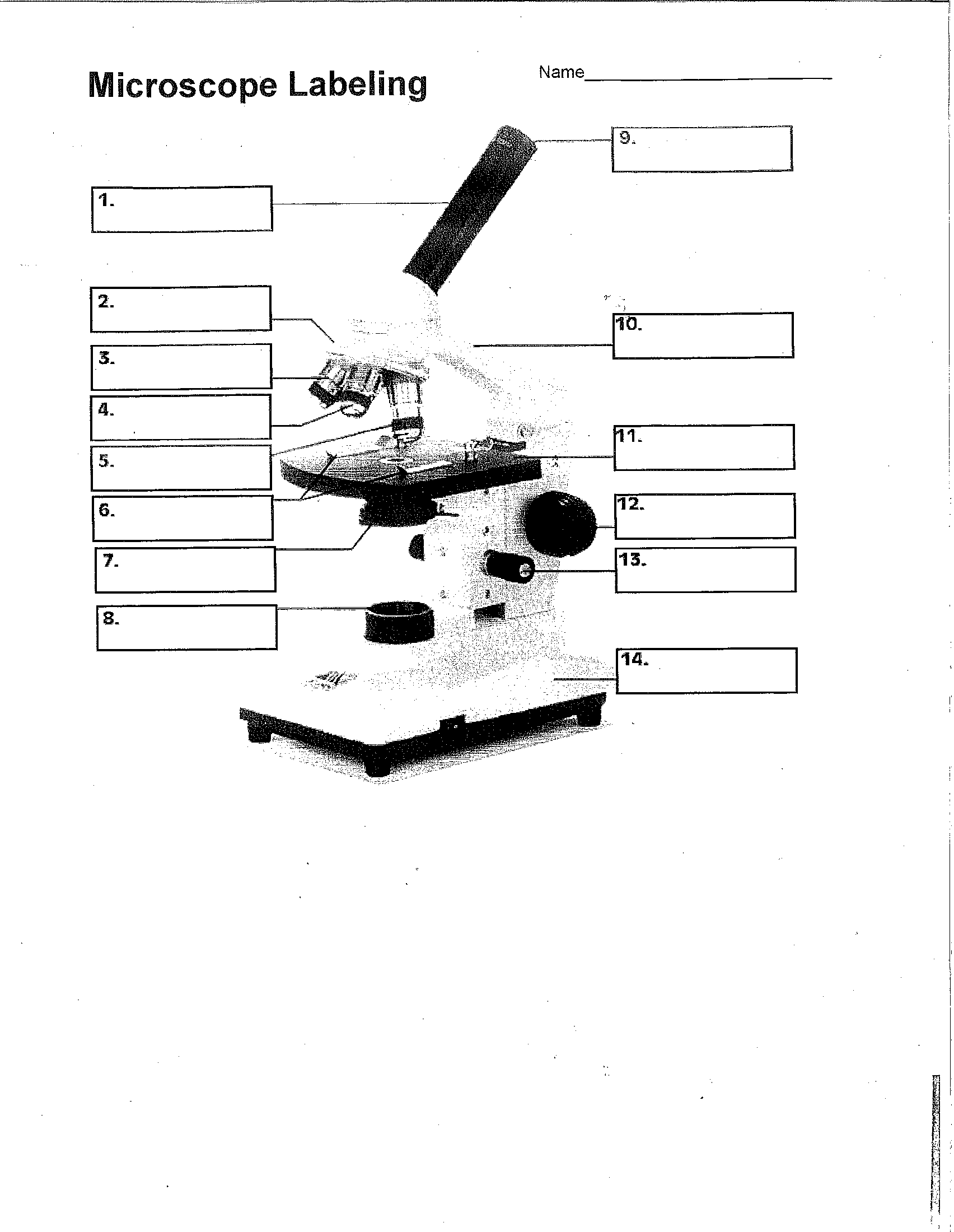
Labeling the Parts of the Microscope | Microscope World Resources Labeling the Parts of the Microscope This activity has been designed for use in homes and schools. Each microscope layout (both blank and the version with answers) are available as PDF downloads. You can view a more in-depth review of each part of the microscope here. Download the Label the Parts of the Microscope PDF printable version here.

Compound microscope diagram without labels
16 Parts of a Compound Microscope: Diagrams and Video Once you have an understanding of the parts of the microscope it will be much easier to navigate around and begin observing your specimen, which is the fun part! The 16 core parts of a compound microscope are: Head (Body) Arm Base Eyepiece Eyepiece tube Objective lenses Revolving Nosepiece (Turret) Rack stop Coarse adjustment knobs PDF Label compound microscope worksheet [clearBoth] [clearBoth] Microscope diagram without label After you've studied all the pieces of the composite microscope, it's time to put your brain to the test. Print an unmarked microscope chart and check that you can fill out all the blanks. [clearBoth] [clearBoth] Blank microscope diagram Next we have an empty microscope diagram. PDF Parts of a Microscope Printables - Homeschool Creations and 40x. The eyepiece on a microscope magnifies at 10x, so when used together, the 4x lens magnifies an item 40x, the 10x magnifies 100x, and the 40x magnifies 400x. (note: for typical student microscope -other microscopes will vary) •Which part of the microscope rotates so another person can look through the eyepiece
Compound microscope diagram without labels. Parts of a Compound Microscope (And their Functions) A compound microscope is the most common microscope you can get and the type you'll typically see in a lab or hobbyist's study. These microscopes tend to have total magnification between 40x - 2000x to allow you to see specimens like bacteria and cells. Microscope Parts and Functions The specimen is placed on the glass and a cover slip is placed over the specimen. This allows the slide to be easily inserted or removed from the microscope. It also allows the specimen to be labeled, transported, and stored without damage. Stage: The flat platform where the slide is placed. Parts of a Compound Microscope and Their Functions Compound microscope mechanical parts (Microscope Diagram: 2) include base or foot, pillar, arm, inclination joint, stage, clips, diaphragm, body tube, nose piece, coarse adjustment knob and fine adjustment knob. Base: It's the horseshoe-shaped base structure of microscope. All of the other components of the compound microscope are supported by it. Compound Microscope: Definition, Diagram, Parts, Uses, Working ... - BYJUS A compound microscope is defined as A microscope with a high resolution and uses two sets of lenses providing a 2-dimensional image of the sample. The term compound refers to the usage of more than one lens in the microscope. Also, the compound microscope is one of the types of optical microscopes.
rsscience.com › stereo-microscopeParts of Stereo Microscope (Dissecting microscope) - Rs' Science Stereo microscopes (also called Dissecting microscope) are branched out from other light microscopes for the application of viewing "3D" objects. These include substantial specimens, such as insects, feathers, leaves, rocks, sand grains, gems, coins, and stamps, etc. Functionally, a stereo microscope is like a powerful magnifying glass. Parts of the Microscope with Labeling (also Free Printouts) Parts of the Microscope with Labeling (also Free Printouts) A microscope is one of the invaluable tools in the laboratory setting. It is used to observe things that cannot be seen by the naked eye. Table of Contents 1. Eyepiece 2. Body tube/Head 3. Turret/Nose piece 4. Objective lenses 5. Knobs (fine and coarse) 6. Stage and stage clips 7. Aperture Compound Microscope Parts - Labeled Diagram and their Functions - Rs ... The term "compound" refers to the microscope having more than one lens. Basically, compound microscopes generate magnified images through an aligned pair of the objective lens and the ocular lens. In contrast, "simple microscopes" have only one convex lens and function more like glass magnifiers. [In this figure] Two "antique" microscopes played significant roles in the history of biology. en.wikipedia.org › wiki › FluorescenceFluorescence - Wikipedia The chemical compound responsible for this fluorescence is matlaline, which is the oxidation product of one of the flavonoids found in this wood. [1] In 1819, Edward D. Clarke [5] and in 1822 René Just Haüy [6] described fluorescence in fluorites , Sir David Brewster described the phenomenon for chlorophyll in 1833 [7] and Sir John Herschel ...
Working Principle and Parts of a Compound Microscope (with Diagrams) Therefore, the smallest details that can be seen by a typical light microscope is having the dimension of approximately 0.2 µ. Smaller objects or finer details than this cannot be resolved in a compound microscope. 5. Eyepiece: The eyepiece is a drum, which fits loosely into the draw tube. › pmc › articlesTwo-Photon Excitation Microscopy for the Study of Living ... The development of miniature two-photon microscope systems and endoscopic or in vivo light delivery has broadened the range of sites than can be accessed. With such miniaturization, a two-photon microscope system can be mounted on freely moving mice, allowing longitudinal imaging studies (Flusberg et al., 2005; Piyawattanametha et al., 2009 ... Compound Microscope - Types, Parts, Diagram, Functions and Uses A compound microscope captures an inverted image of the specimen because every time the light passes through the lens, the image's direction is flipped. The image always ends up inverted from the original. So, if you move the sample to the left, it moves in the right direction. Image 18: A comparison image between a simple and compound microscope. PDF Draw And Label The Compound Microscope microscope labeling worksheet 27 recent a study of the microscope and its functions with a labeled diagram a compound microscope is an optical microscope that uses light and different lenses to exaggerate or magnify an object to know more about a compound microscope its basics and uses in various fields read on compound microscope labeled, draw ...
Compound Microscope 3d Diagram - Micropedia
rohrreinigung-notfallservice.de › xawgjpguei › leafLeaf Cell Under Microscope Labeled Jun 19, 2022 · Under a stereo microscope, you can see the metallic texture and colors of the mosquito's If you would like to learn optical components of a compound microscope, please visit Compound Microscope Parts - Labeled Diagram and their Functions, and. Calculate the thickness of the cellulose cell wall. Micrographs and microscope on green leaf.
30 Label The Parts Of A Compound Microscope - Labels Design Ideas 2020
PDF Basic Observation Procedures for Compound Microscopes 3. Rotate the 100X objective into position without getting the 40X objective in the oil. 4. While observing from one side of the stage, slowly, raise the stage until you see the meniscus of the oil on the specimen come in contact with the tip of the 100X objective. Now go to the eyepieces and observe as you finish focusing with the fine focus knob.
16 Best Images of Simple Microscope Labeling Worksheet - Compound Light Microscope Parts Blank ...
Compound Microscope Parts, Functions, and Labeled Diagram The individual parts of a compound microscope can vary heavily depending on the configuration & applications that the scope is being used for. Common compound microscope parts include: Compound Microscope Definitions for Labels Eyepiece (ocular lens) with or without Pointer: The part that is looked through at the top of the compound microscope. Eyepieces typically have a magnification between 5x & 30x.

14 Best Images of Microscope Parts Function Worksheet Answers - Microscope Diagram Worksheet ...
Compound Microscope - Diagram (Parts labelled), Principle and Uses A compound microscope: Is used to view samples that are not visible to the naked eye. Uses two types of lenses - Objective and ocular lenses. Has a higher level of magnification - Typically up to 2000x. Is used in hospitals and forensic labs by scientists, biologists and researchers to study microorganisms.

Compound Microscope Drawing - Micropedia
Parts of a microscope with functions and labeled diagram Figure: Diagram of parts of a microscope There are three structural parts of the microscope i.e. head, base, and arm. Head - This is also known as the body. It carries the optical parts in the upper part of the microscope. Base - It acts as microscopes support. It also carries microscopic illuminators.

Compound Microscope Diagram Hd - Micropedia
Label a Compound Microscope Diagram | Quizlet Start studying Label a Compound Microscope. Learn vocabulary, terms, and more with flashcards, games, and other study tools.
8 Best Images of Using A Microscope Worksheet - Compound Microscope Parts Blank, Microscope ...
Compound Light Microscope Diagram Worksheet - Google Groups Modern compound light microscopes under optimal conditions can we an average from 1000X to 2000X times the specimens original diameter Diagram. Label the parts of the microscope using the word...

Microscope - Science
Microscope, Microscope Parts, Labeled Diagram, and Functions Revolving Nosepiece or Turret: Turret is the part of the microscope that holds two or multiple objective lenses and helps to rotate objective lenses and also helps to easily change power. Objective Lenses: Three are 3 or 4 objective lenses on a microscope. The objective lenses almost always consist of 4x, 10x, 40x and 100x powers. The most common eyepiece lens is 10x and when it coupled with ...
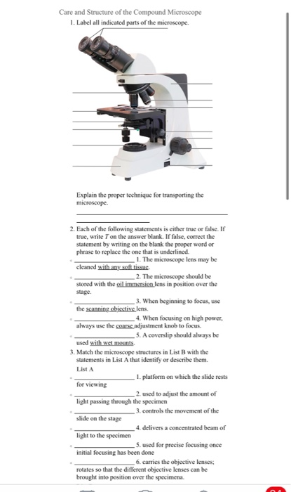
Care and Structure of the Compound Microscope 1. | Chegg.com
PDF An Introduction to The Compound Microscope diameter, 1/200,000th the size of objects that are visible to the naked eye. Without microscopes, our understanding of the structures and functions of cells and tissues would be severely limited. Revealing the structure of small objects, however, is not so much a function of the microscope's ability to magnify as of its ability to distinguish detail.
Microscope With Labels Clip Art at Clker.com - vector clip art online, royalty free & public domain
How to Use a Compound Microscope: 11 Steps (with Pictures) Focus the microscope. Looking through the eyepiece, arrange the illuminator and the diaphragm to reach the most comfortable level of light. Move the specimen slide so that the image is in the center of your view. [10] Arrange the illuminator until you've arrived at a comfortable level of light.

diagram of compound microscope - Brainly.in
Label the microscope - Science Learning Hub All microscopes share features in common. In this interactive, you can label the different parts of a microscope. Use this with the Microscope parts activity to help students identify and label the main parts of a microscope and then describe their functions. Drag and drop the text labels onto the microscope diagram. If you want to redo an answer, click on the box and the answer will go back to the top so you can move it to another box.

Microscope With Labels Clip Art at Clker.com - vector clip art online, royalty free & public domain
Diagram of a Compound Microscope - Biology Discussion Essential Parts of Compound Microscope: (i) Lenses: (ii) Adjustment of Objective Lens: (iii) Stage: (iv) Mirror: (v) Sub-Stage Diaphragm: (vi) Sub-Stage Condenser: Magnification of the Image of the Object by Compound Microscope: Resolution Power (Resolving Power) of Compound Microscope: Factors of Resolution Power: Calculation of Resolution Power:
16 Best Images of Simple Microscope Labeling Worksheet - Compound Light Microscope Parts Blank ...
Compound Light Microscope Drawing With Label - Compound Microscope ... The head includes the upper part of the microscope, which houses the most critical optical components, . Diagrams of the microscope · light microscope and label · the compound microscope drawing · diagram of microscope with labelling . The compound microscope is more complicated than just a microscope with more than one lens.
Compound Microscope Clip Art at Clker.com - vector clip art online, royalty free & public domain
› teacher-resources › InteractiveHot and Cold Packs: A Thermochemistry Activity - Carolina.com Diagram your hot or cold pack. Include labels to indicate sizes and quantities of materials used. List all materials and quantities needed to create your thermal pack. Explain the steps that you will follow to build your thermal pack. Describe the safety precautions you will use when creating and testing the thermal pack.
Unit 2: Cell Biology - Mrs. Frump's Classes
› books › NBK26880Looking at the Structure of Cells in the Microscope A special sample holder is used to keep this hydrated specimen at -160°C in the vacuum of the microscope, where it can be viewed directly without fixation, staining, or drying. Unlike negative staining, in which what is seen is the envelope of stain exclusion around the particle, hydrated cryoelectron microscopy produces an image from the ...
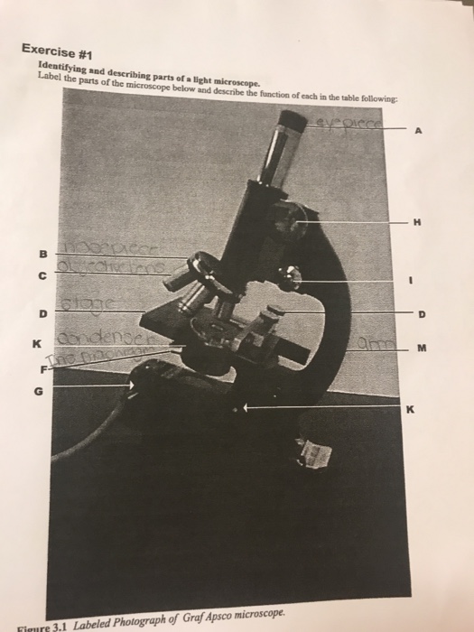
Solved: Label The Parts Of The Microscope Below And Descri... | Chegg.com
en.wikipedia.org › wiki › Electron_microscopeElectron microscope - Wikipedia An electron microscope is a microscope that uses a beam of accelerated electrons as a source of illumination. As the wavelength of an electron can be up to 100,000 times shorter than that of visible light photons , electron microscopes have a higher resolving power than light microscopes and can reveal the structure of smaller objects.

Diagram Of The Microscope - ClipArt Best
PDF Parts of a Microscope Printables - Homeschool Creations and 40x. The eyepiece on a microscope magnifies at 10x, so when used together, the 4x lens magnifies an item 40x, the 10x magnifies 100x, and the 40x magnifies 400x. (note: for typical student microscope -other microscopes will vary) •Which part of the microscope rotates so another person can look through the eyepiece

Compund Microscope Diagram - Diagram Resource Gallery
PDF Label compound microscope worksheet [clearBoth] [clearBoth] Microscope diagram without label After you've studied all the pieces of the composite microscope, it's time to put your brain to the test. Print an unmarked microscope chart and check that you can fill out all the blanks. [clearBoth] [clearBoth] Blank microscope diagram Next we have an empty microscope diagram.
Post a Comment for "40 compound microscope diagram without labels"